
AI for DVT Ultrasound
You're better off doing it yourself.
There’s a lot of AI out there that’s technology looking for a problem in medicine to solve – see: LLMs. This is the lovely opposite, a clinical and resource problem requiring a technology solution.
But, it might take until version 2.0.
It’s a simple application of AI image processing to guide evaluation for deep venous thrombosis using a two-site compression technique augmented by machine learning. In this trial comparing “AutoDVT” to sonographer gold standard, the authors analyzed 294 patients in a couple different ways, and these two tables convey the bulk of the findings:
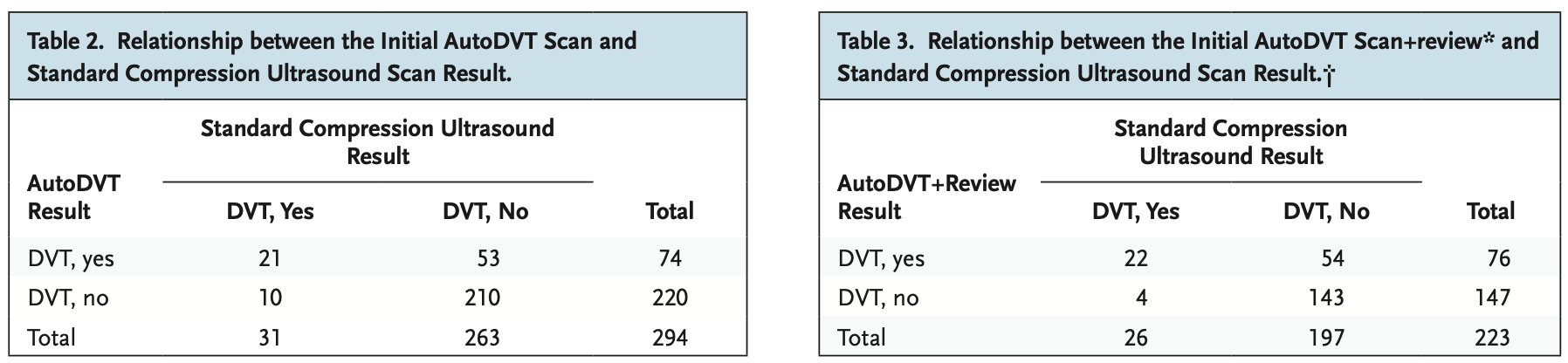
Table 2 shows the performance of AutoDVT in isolation – a sensitivity of 68% and specificity of 80%. Unusable.
Table 3 is an interesting secondary analysis – using a radiologist review images captured by the AutoDVT guided system. This performs a sort of test to see whether the problem was poor image quality being captured by these non-sonographer users, or whether the problem was localized to the diagnostic image processing. In this analysis, a “perfect” interpretation of images had a sensitivity of 85% and a specificity of 73%. Also unusable – and indicates there are issues with both guided image acquisition and interpretation.
A worthy unmet need for resource-limited settings, and I expect these test characteristics are likely to be surpassed in future iterations.